Understanding Animal Cell Structure

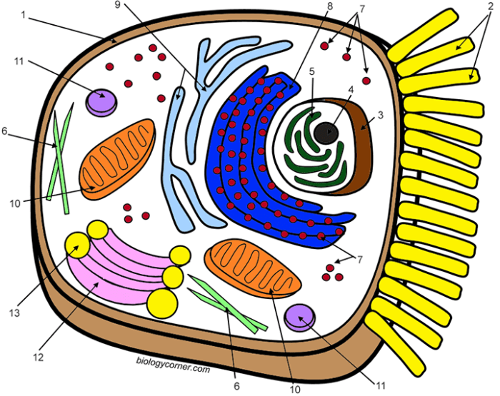
Animal cell coloring biology corner answer key – Animal cells are the fundamental building blocks of animals, exhibiting a complex internal organization crucial for their survival and function. Understanding their structure is essential to grasping the intricacies of life itself. This section will explore the major components of an animal cell, their individual roles, and their interdependencies.
The Cell Membrane: Maintaining Cell Integrity
The cell membrane, also known as the plasma membrane, is a selectively permeable barrier that encloses the entire cell. It’s a phospholipid bilayer, meaning it consists of two layers of phospholipid molecules arranged with their hydrophilic (water-loving) heads facing outward and their hydrophobic (water-fearing) tails facing inward. Embedded within this bilayer are various proteins that perform diverse functions, including transport of molecules, cell signaling, and cell adhesion.
The cell membrane’s selective permeability allows the cell to regulate the passage of substances in and out, maintaining a stable internal environment crucial for cellular processes. This regulation prevents the uncontrolled influx or efflux of essential molecules, ensuring the cell’s survival. For instance, it controls the movement of ions like sodium and potassium, essential for nerve impulse transmission.
Major Organelles and Their Functions
Animal cells contain various organelles, each with specialized functions contributing to the overall cellular activity. These organelles work in coordination to maintain the cell’s life processes.
| Organelle | Location | Function | Typical Color |
|---|---|---|---|
| Cell Membrane | Outer boundary of the cell | Regulates passage of substances; maintains cell integrity | Light brown/Beige |
| Nucleus | Usually centrally located | Contains genetic material (DNA); controls cell activities | Purple |
| Mitochondria | Scattered throughout the cytoplasm | Generates energy (ATP) through cellular respiration | Red |
| Ribosomes | Free-floating in cytoplasm or attached to ER | Synthesize proteins | Dark blue/Black |
| Endoplasmic Reticulum (ER) | Network of membranes throughout cytoplasm | Synthesizes and transports proteins and lipids | Light blue |
| Golgi Apparatus | Near the nucleus | Modifies, sorts, and packages proteins and lipids | Yellow |
| Lysosomes | Throughout cytoplasm | Break down waste materials and cellular debris | Green |
| Cytoskeleton | Throughout cytoplasm | Provides structural support and facilitates movement | Light gray |
Comparison of Nucleus, Mitochondria, and Ribosomes, Animal cell coloring biology corner answer key
The nucleus, mitochondria, and ribosomes are crucial organelles with distinct structures and functions. The nucleus, a large, membrane-bound organelle, houses the cell’s genetic material (DNA) and controls gene expression, directing cellular activities. Mitochondria, also membrane-bound, are the powerhouses of the cell, generating ATP through cellular respiration. Ribosomes, much smaller and lacking a membrane, are the protein synthesis machinery of the cell, translating genetic information into functional proteins.
The nucleus dictates the production of proteins, the mitochondria provide the energy for protein synthesis, and the ribosomes carry out the actual synthesis. This interconnectedness highlights the coordinated function of these essential organelles.
The Biology Corner Resource: Animal Cell Coloring Biology Corner Answer Key

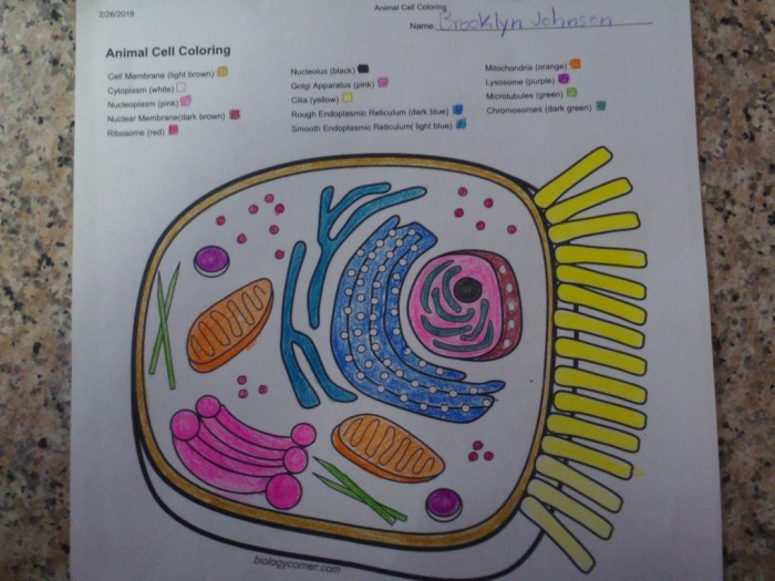
The Biology Corner’s animal cell coloring worksheet serves as a valuable tool for introducing students to the fundamental structures of animal cells. It leverages a multi-sensory approach to learning, combining visual representation with active engagement. This approach is particularly effective for younger learners or those who benefit from hands-on activities.
Learning Objectives Addressed by the Worksheet
The worksheet likely aims to achieve several key learning objectives. Students are expected to learn the names and functions of major organelles within an animal cell, such as the nucleus, cytoplasm, mitochondria, ribosomes, and cell membrane. Furthermore, the activity promotes understanding of the cell’s overall structure and the relationships between its different components. Finally, it reinforces visual recognition and memorization of these organelles, improving recall and comprehension.
Pedagogical Approach of the Worksheet
The Biology Corner worksheet employs a primarily visual learning approach. The coloring activity itself engages students actively in the learning process, reinforcing the visual information through kinesthetic engagement. This active recall, through the process of identifying and coloring specific organelles, aids in memory consolidation. The act of labeling parts further strengthens the connection between the visual representation and the terminology associated with each organelle.
This combination of visual and kinesthetic learning styles caters to diverse learning preferences.
Potential Challenges Students Might Face
Students might encounter challenges related to visual discrimination of similar-looking organelles, particularly under a microscope. Differentiating between the endoplasmic reticulum and the Golgi apparatus, for example, can be difficult. Some students may also struggle with memorizing the functions of each organelle, requiring additional reinforcement. Finally, accurately labeling the structures on the diagram requires a degree of fine motor skill and careful attention to detail.
Supplementary Activity to Enhance Understanding
To further enhance understanding after completing the coloring worksheet, a 3D model construction activity could be beneficial. Students could use readily available materials like clay, play-doh, or even recycled materials to build a three-dimensional representation of an animal cell. This activity would allow for a more tactile and spatial understanding of the relative sizes and positions of different organelles within the cell.
Each organelle could be represented by a different color or texture, further reinforcing visual learning. The construction process would also encourage collaborative learning and problem-solving skills.
The intricate detail of the animal cell coloring biology corner answer key felt strangely satisfying, a quiet triumph over the complexities of cellular structure. It reminded me of simpler times, coloring pictures of adorable farm animals – you can find some wonderful options at free farm animal coloring sheets – before the pressures of biology exams set in.
Returning to the answer key, I felt a renewed sense of calm, ready to tackle the next microscopic challenge.


