Comparative Analysis with Real Animal Cells

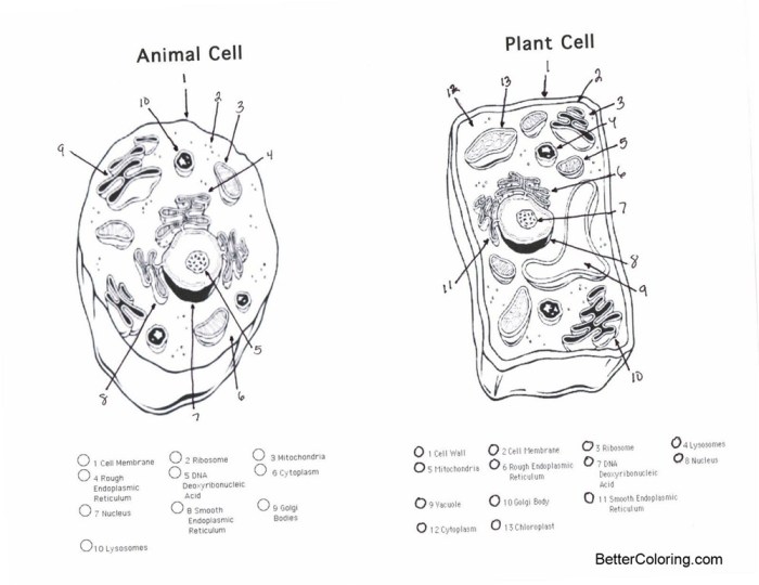
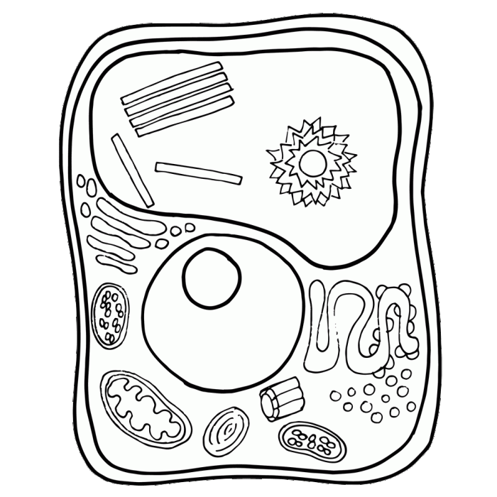
Animal cell coloring page.jpg – A coloring page depicting an animal cell provides a simplified, visually appealing representation of a complex biological structure. However, it’s crucial to understand the limitations of this simplified model when compared to the intricate reality observed under a microscope. This comparison highlights the discrepancies between the illustrative and the actual biological structures, emphasizing the need for diverse learning resources for a comprehensive understanding.Comparing the coloring page’s depiction to a microscopic image reveals both similarities and differences.
Similarities often include the presence of a nucleus (generally centrally located in both), a cell membrane (represented as a boundary in both), and possibly a representation of cytoplasm (the filling of the cell). However, the coloring page lacks the detail and complexity found in a real animal cell image.
Discrepancies Between Simplified and Actual Animal Cell Representations, Animal cell coloring page.jpg
The simplified nature of a coloring page necessitates a discussion of its limitations. Relying solely on a coloring page for understanding animal cell structure is inadequate because it omits crucial details of cellular complexity and function. Additional resources, such as textbooks, microscopic images, and interactive simulations, are necessary to build a complete understanding. These resources provide the depth and detail absent from a two-dimensional, simplified drawing.
While the “animal cell coloring page.jpg” offers a fascinating glimpse into the microscopic world, a different kind of adorable awaits with the charming illustrations found at animal baby cute unicorn coloring pages. These whimsical designs provide a delightful contrast to the intricate detail of the cell structure, reminding us that the beauty of biology extends across vastly different scales.
Returning to the “animal cell coloring page.jpg,” we can appreciate the complexity of even the smallest life forms.
Key Differences Between Coloring Page and Microscopic Images
A list of five key differences helps to illustrate the limitations of the coloring page as a sole learning tool. The following points highlight the significant gap between the simplified representation and the true complexity of an animal cell:
- Organelle Detail: Coloring pages typically show only a few major organelles (like the nucleus and sometimes the cell membrane). Microscopic images reveal a multitude of organelles, each with unique structures and functions, such as mitochondria, endoplasmic reticulum, Golgi apparatus, lysosomes, and ribosomes, which are often omitted or highly simplified in coloring pages.
- Organelle Size and Shape: The relative sizes and shapes of organelles are often inaccurate in coloring pages. Microscopic images show the precise dimensions and forms of these structures, which vary greatly depending on the cell type and its current activity.
- Cytoplasmic Detail: A coloring page usually depicts cytoplasm as a homogenous substance. In contrast, microscopic images show a dynamic environment with various inclusions, cytoskeletal elements (microtubules, microfilaments), and the movement of molecules and organelles.
- Membrane Structure: The cell membrane is often represented as a simple line in a coloring page. Microscopic images, however, show a complex phospholipid bilayer with embedded proteins, illustrating the membrane’s role in selective permeability and cellular communication.
- Three-Dimensional Structure: Coloring pages are two-dimensional representations, failing to capture the three-dimensional nature of a real animal cell. Microscopic images, even in two dimensions, offer a better sense of the spatial arrangement of organelles within the cell, showing the complex interplay between different structures.
Creative Extensions and Adaptations

This section explores alternative designs for an animal cell coloring page, pedagogical approaches for using the page, and a creative narrative incorporating the cell’s components. These extensions aim to enhance learning and engagement with cell biology concepts.
Alternative Animal Cell Coloring Page Designs
Three alternative designs offer different perspectives on animal cell structure and function. The first focuses on the dynamic processes within the cell, the second emphasizes the detailed structure of individual organelles, and the third highlights the crucial role of the cell membrane.
- Design 1: Cell Processes in Action: This design depicts the cell actively engaged in various processes. Vesicles are shown moving between the endoplasmic reticulum and Golgi apparatus, highlighting protein synthesis and transport. Mitochondria are illustrated actively producing ATP, with arrows indicating energy flow. Lysosomes are depicted breaking down waste materials. The nucleus is shown undergoing transcription and mRNA leaving the nuclear pores.
The overall visual is dynamic, showcasing the cell’s constant activity.
- Design 2: Organelle Detail: This design provides a magnified view of individual organelles. Each organelle is depicted in greater detail, showing internal structures like the cristae of mitochondria or the ribosomes on the rough endoplasmic reticulum. Labels clearly identify each organelle and its key features. This design emphasizes the intricate organization within the cell.
- Design 3: Cell Membrane Focus: This design emphasizes the cell membrane and its interactions with the external environment. The phospholipid bilayer is clearly illustrated, along with embedded proteins involved in transport, cell signaling, and cell adhesion. Extracellular components, such as the extracellular matrix, are also shown, highlighting the membrane’s role in maintaining cell integrity and communication.
Activities for Teaching Organelle Functions
Using the coloring page, students can learn the functions of different organelles through interactive activities. For example, students can color-code organelles based on their function (e.g., energy production, protein synthesis, waste removal). They can then create diagrams or flowcharts illustrating how organelles work together to maintain cell function.
- Mitochondria: Students can color the mitochondria a vibrant red to represent energy production, and label them with the process of cellular respiration.
- Ribosomes: Students can color ribosomes blue to represent protein synthesis and draw arrows to show the movement of mRNA and proteins.
- Golgi Apparatus: Students can color the Golgi apparatus green to represent processing and packaging of proteins, showing the movement of vesicles.
- Lysosomes: Students can color lysosomes purple to represent waste breakdown and show them engulfing cellular debris.
A Cell’s Tale
Once upon a time, in the bustling metropolis of the human body, lived a plucky animal cell named Celia. Celia was a vibrant, bustling place, a miniature city teeming with activity. Her nucleus, the city hall, held the master blueprints for all her functions.
The mitochondria, Celia’s power plants, tirelessly generated energy, keeping her humming with life. The endoplasmic reticulum, her vast network of highways, transported essential materials throughout her cytoplasm.
The Golgi apparatus, Celia’s efficient postal service, packaged and shipped proteins to their designated destinations. The lysosomes, her diligent sanitation department, diligently cleaned up cellular waste.
One day, a foreign invader, a virus, tried to breach Celia’s defenses. But her cell membrane, her sturdy city walls, repelled the attack. Celia, with the help of her organelles, successfully defended herself, proving the resilience and power of even the smallest cell.
Quick FAQs: Animal Cell Coloring Page.jpg
What are some common mistakes found in student’s coloring pages of animal cells?
Common mistakes include incorrectly labeling organelles, misrepresenting their shapes and sizes, and omitting key structures entirely.
How can I adapt the coloring page for students with different learning styles?
Adaptations include providing tactile versions for visually impaired students, offering audio descriptions, and incorporating kinesthetic activities.
Are there any online resources that complement the coloring page?
Yes, many interactive websites and educational videos provide detailed information about animal cells, offering supplementary learning materials.
What are some alternative ways to use this coloring page beyond coloring?
Students could use it as a template for creating 3D models, designing diagrams, or writing short stories about the cell’s activities.


