Introduction to Animal Cell Structure
Animal cell labeling and coloring – Animal cells are the fundamental building blocks of animals, exhibiting a complex internal organization crucial for their diverse functions. Understanding their structure is key to comprehending the processes of life itself. These cells, unlike plant cells, lack a rigid cell wall and chloroplasts, resulting in a more flexible and adaptable structure.The animal cell is characterized by a variety of membrane-bound organelles, each with a specific role in maintaining cellular homeostasis and carrying out essential life processes.
These organelles work in concert, creating a highly organized and efficient system.
Cell Membrane
The cell membrane, also known as the plasma membrane, is a selectively permeable barrier that encloses the cell’s contents. It’s composed primarily of a phospholipid bilayer, a double layer of lipid molecules with hydrophilic (water-loving) heads facing outwards and hydrophobic (water-fearing) tails facing inwards. Embedded within this bilayer are various proteins that perform diverse functions, including transport of molecules, cell signaling, and cell adhesion.
The cell membrane regulates the passage of substances into and out of the cell, maintaining a stable internal environment. This selective permeability is crucial for maintaining the cell’s internal balance and protecting it from harmful substances. The fluidity of the membrane allows for movement of its components, enabling adaptation and response to changes in the cellular environment.
For example, receptor proteins on the membrane can bind to specific signaling molecules, triggering intracellular responses.
Cytoplasm, Animal cell labeling and coloring
The cytoplasm is the jelly-like substance that fills the cell between the cell membrane and the nucleus. It’s a complex mixture of water, salts, and various organic molecules, including enzymes, nutrients, and waste products. The cytoplasm serves as a medium for various cellular processes, providing a location for metabolic reactions and the movement of organelles. The cytoskeleton, a network of protein filaments within the cytoplasm, provides structural support and facilitates intracellular transport.
Nucleus
The nucleus is the control center of the cell, containing the cell’s genetic material – DNA (deoxyribonucleic acid). DNA is organized into chromosomes, which carry the instructions for building and maintaining the cell. The nucleus is surrounded by a double membrane called the nuclear envelope, which contains nuclear pores that regulate the transport of molecules between the nucleus and the cytoplasm.
Within the nucleus, the nucleolus is a dense region where ribosomes, essential for protein synthesis, are assembled.
Ribosomes
Ribosomes are small organelles responsible for protein synthesis. They are either free-floating in the cytoplasm or attached to the endoplasmic reticulum. Ribosomes translate the genetic code from messenger RNA (mRNA) into polypeptide chains, which fold into functional proteins. These proteins are essential for virtually all cellular processes.
Endoplasmic Reticulum (ER)
The endoplasmic reticulum (ER) is a network of interconnected membranes extending throughout the cytoplasm. There are two types: rough ER and smooth ER. Rough ER, studded with ribosomes, is involved in protein synthesis and modification. Smooth ER, lacking ribosomes, plays a role in lipid synthesis, detoxification, and calcium storage.
Golgi Apparatus
The Golgi apparatus, also known as the Golgi complex, is a stack of flattened, membrane-bound sacs. It receives proteins and lipids from the ER, modifies them, sorts them, and packages them into vesicles for transport to other parts of the cell or for secretion outside the cell. Think of it as the cell’s post office.
Mitochondria
Mitochondria are the powerhouses of the cell, responsible for generating most of the cell’s energy in the form of ATP (adenosine triphosphate) through cellular respiration. They have a double membrane structure, with the inner membrane folded into cristae, increasing the surface area for ATP production. Mitochondria possess their own DNA and ribosomes, suggesting an endosymbiotic origin.
Lysosomes
Lysosomes are membrane-bound organelles containing digestive enzymes that break down waste materials, cellular debris, and ingested particles. They are involved in recycling cellular components and defending the cell against pathogens.
Labeling Animal Cell Diagrams
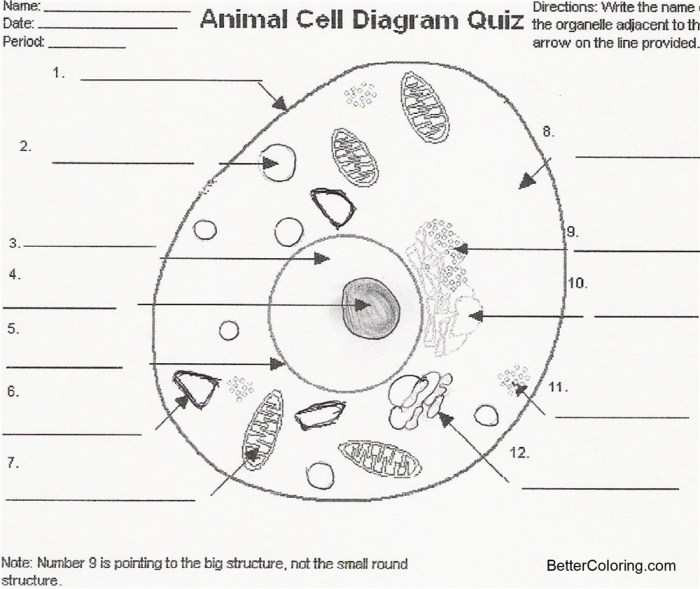
Accurately labeling diagrams of animal cells is crucial for understanding their complex structure and the functions of their various components. Clear and precise labeling allows for effective communication of scientific information and facilitates a deeper understanding of cellular processes. This section will guide you through the process of labeling an animal cell diagram, emphasizing the importance of accuracy and consistency.
Creating a labeled diagram involves identifying each organelle, understanding its function, and correctly placing it within the cell’s structure. Consistent color-coding helps to visually organize the information and improve comprehension.
Animal Cell Organelle Information
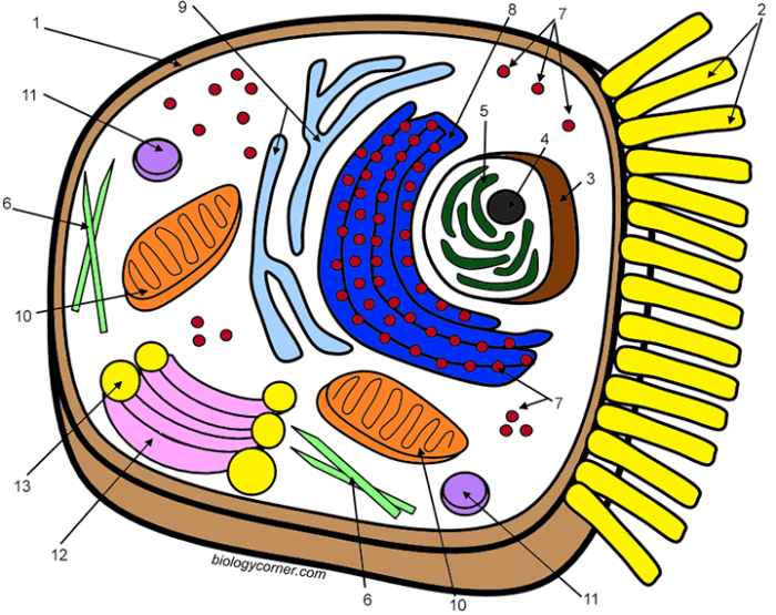
The following table provides a comprehensive list of major animal cell organelles, their functions, typical locations within a cell diagram, and suggested color codes for visual representation. Remember, these color codes are suggestions; you can adapt them to your preferences while maintaining consistency within your diagram.
| Organelle | Function | Location in Diagram | Color Code |
|---|---|---|---|
| Nucleus | Contains genetic material (DNA); controls cell activities. | Central, usually largest organelle. | Dark Purple |
| Nucleolus | Produces ribosomes. | Within the nucleus. | Light Purple |
| Ribosomes | Synthesize proteins. | Free-floating in cytoplasm or attached to ER. | Dark Blue |
| Endoplasmic Reticulum (ER) | Rough ER: protein synthesis and transport; Smooth ER: lipid synthesis and detoxification. | Network of membranes throughout cytoplasm. | Light Blue (Rough ER), Light Green (Smooth ER) |
| Golgi Apparatus (Golgi Body) | Processes and packages proteins and lipids. | Near the nucleus, often stacked membrane sacs. | Yellow |
| Mitochondria | Produce ATP (energy) through cellular respiration. | Scattered throughout cytoplasm. | Red |
| Lysosomes | Break down waste materials and cellular debris. | Scattered throughout cytoplasm. | Orange |
| Cytoplasm | Gel-like substance filling the cell; site of many metabolic reactions. | Fills the space between organelles. | Light Tan |
| Cell Membrane | Regulates what enters and leaves the cell. | Outer boundary of the cell. | Light Brown |
| Centrioles | Involved in cell division. | Near the nucleus, usually in pairs. | Dark Green |
Diagram of an Animal Cell with Labels
Imagine a circular cell. The nucleus, a large dark purple circle, sits centrally. Within it, a smaller light purple circle represents the nucleolus. The cytoplasm, a light tan background, fills the cell. Scattered throughout the cytoplasm are numerous small dark blue dots (ribosomes), larger red ovals (mitochondria), and smaller orange circles (lysosomes).
A network of light blue (rough ER) and light green (smooth ER) tubules extends throughout the cytoplasm. Near the nucleus, a stack of yellow flattened sacs represents the Golgi apparatus. A light brown line delineates the cell membrane. Near the nucleus, a pair of dark green cylindrical structures represent the centrioles. A key at the bottom of the diagram would list each organelle with its corresponding color.
Importance of Accurate Labeling
Accurate labeling is paramount in cell diagrams. Inaccurate or missing labels can lead to misinterpretations of the cell’s structure and function. Precise labeling ensures clear communication of scientific information, allowing for effective collaboration and understanding among researchers and students alike. Furthermore, it promotes critical thinking and attention to detail, essential skills in any scientific endeavor. For instance, mislabeling a mitochondrion as a lysosome would significantly alter the understanding of the cell’s energy production and waste disposal processes.
The consequences of inaccurate labeling can be far-reaching, potentially affecting research outcomes and educational understanding.
Illustrating Specific Organelles

Understanding the function of an animal cell requires a close examination of its individual components, or organelles. Each organelle plays a crucial role in maintaining the cell’s overall health and function. This section will focus on three particularly important organelles: the mitochondria, lysosomes, and the nucleus.Mitochondria, lysosomes, and the nucleus are significantly different in both size and appearance, reflecting their distinct roles within the cell.
Their internal structures are also highly specialized, contributing to their unique functionalities.
Understanding animal cell labeling and coloring helps visualize the intricate structures within. This detailed process is surprisingly similar to the fun of coloring, like those found on a delightful animal babies coloring page , where you bring adorable creatures to life with color. Both activities encourage observation and precision, developing valuable skills applicable to scientific study and creative expression.
Returning to the scientific realm, accurate animal cell labeling is crucial for accurate biological understanding.
Mitochondrial Structure and Function
Mitochondria are often referred to as the “powerhouses” of the cell because they are responsible for generating most of the cell’s supply of adenosine triphosphate (ATP), the primary energy currency. Their structure is highly organized to facilitate this energy production. Imagine a mitochondrion as a bean-shaped organelle with a double membrane. The outer membrane is smooth, while the inner membrane is highly folded into cristae, creating a large surface area for the electron transport chain, a key process in ATP synthesis.
Within the inner membrane is the mitochondrial matrix, a gel-like substance containing enzymes and other molecules involved in cellular respiration.Illustration: A mitochondrion is depicted as a bean-shaped structure. The outer membrane is represented as a smooth, continuous line, while the inner membrane is shown as numerous folds (cristae) extending into the interior. The space enclosed by the inner membrane is the matrix, which appears as a lightly shaded area containing small dots representing enzymes and other molecules.
A caption could read: “Mitochondrion: The bean-shaped organelle with folded inner membranes (cristae) maximizing surface area for ATP production.”
Lysosomal Structure and Function
Lysosomes are membrane-bound organelles containing hydrolytic enzymes, which are crucial for breaking down various substances within the cell. They act as the cell’s recycling and waste disposal system. These organelles are typically spherical in shape and are relatively small compared to other organelles like the nucleus or mitochondria. Their single membrane encloses a highly acidic environment, optimal for the activity of the hydrolytic enzymes.
These enzymes break down various materials, including cellular debris, worn-out organelles, and ingested pathogens.Illustration: A lysosome is depicted as a small, spherical vesicle with a single, clearly defined membrane. The interior is shaded to represent the acidic environment and contains small, irregularly shaped shapes representing hydrolytic enzymes. A caption could read: “Lysosome: A spherical vesicle containing hydrolytic enzymes for cellular waste breakdown and recycling.”
Nuclear Structure and Function
The nucleus is the largest and arguably the most important organelle in most animal cells. It acts as the cell’s control center, housing the cell’s genetic material (DNA) and regulating gene expression. The nucleus is a roughly spherical structure enclosed by a double membrane, the nuclear envelope, which is perforated by nuclear pores that regulate the transport of molecules between the nucleus and the cytoplasm.
Inside the nucleus, DNA is organized into chromosomes, and a distinct region called the nucleolus is responsible for ribosome synthesis.Illustration: The nucleus is depicted as a large, spherical structure with a double membrane (nuclear envelope) showing numerous small pores. The interior contains a darker, irregularly shaped region representing the nucleolus and a diffuse, lightly shaded area representing chromatin (DNA).
A caption could read: “Nucleus: The control center of the cell, containing DNA and the nucleolus for ribosome production.”
Creating a Comprehensive Animal Cell Model
Building a three-dimensional model of an animal cell is a fantastic way to solidify your understanding of its intricate structure and the functions of its various organelles. This hands-on approach allows for a deeper appreciation of the cell’s complexity beyond simply looking at diagrams. By carefully selecting materials and following a structured approach, you can create a visually engaging and informative model.Creating a three-dimensional model involves selecting appropriate materials to represent the different organelles and carefully assembling them to accurately reflect the cell’s structure.
The choice of materials depends on factors such as availability, ease of use, and visual clarity.
Materials and Tools for Animal Cell Model Construction
The materials you choose will significantly impact the final look and feel of your model. Consider using materials that are easily manipulated and readily available. Furthermore, the tools selected should be appropriate for handling the chosen materials.
- Base: A styrofoam ball or a similarly shaped object provides a solid foundation for your model. Its size will dictate the overall scale of your cell.
- Nucleus: A smaller styrofoam ball, painted dark purple or brown, will represent the cell’s nucleus. You can add a smaller, lighter-colored ball inside to represent the nucleolus.
- Mitochondria: Use small, oblong-shaped candies (like jelly beans) or appropriately sized beads, colored dark red or brown, to depict mitochondria. These can be attached to the surface of the main ball.
- Endoplasmic Reticulum (ER): Thin strips of construction paper, folded and arranged to resemble a network, can effectively represent the ER. Use different colors for the rough ER (studded with ribosomes) and smooth ER.
- Ribosomes: Small sprinkles or beads can be used to represent ribosomes. These can be attached to the rough ER strips.
- Golgi Apparatus: Several flat, stacked pieces of cardboard or construction paper, slightly curved, can represent the flattened sacs of the Golgi apparatus. These can be positioned near the nucleus.
- Lysosomes: Small, round candies or beads, colored dark blue or green, can represent lysosomes. These are usually scattered throughout the cytoplasm.
- Vacuoles: Small, clear plastic bags filled with colored water can effectively represent vacuoles. Their size can vary depending on the type of animal cell being modeled.
- Cytoskeleton: Thin strands of yarn or pipe cleaners can be used to represent the cytoskeleton, which provides structural support. These are interwoven throughout the model.
- Cell Membrane: A clear plastic bag or cellophane wrap can be used to create a “skin” for the cell, representing the cell membrane. This can be secured around the styrofoam ball.
Beyond the materials listed above, you will need basic tools like scissors, glue, paint, markers, and toothpicks or pins to secure the different components. Remember safety precautions when using sharp objects.
Step-by-Step Guide to Animal Cell Model Construction
Constructing the model requires a methodical approach to ensure accurate representation of the organelles and their spatial relationships. Carefully assembling the components will result in a more accurate and visually appealing model.
- Prepare the Base: Begin by painting your styrofoam ball to represent the cytoplasm (a light beige or pale yellow works well).
- Add the Nucleus: Attach the smaller styrofoam ball (the nucleus) to the larger ball using glue or toothpicks.
- Position Organelles: Carefully glue or pin the remaining organelles (mitochondria, Golgi apparatus, ER, lysosomes, and vacuoles) to the surface of the larger ball, ensuring they are positioned accurately relative to each other and the nucleus.
- Integrate the Cytoskeleton: Gently weave the yarn or pipe cleaners representing the cytoskeleton throughout the model.
- Complete the Cell Membrane: Carefully secure the clear plastic bag or cellophane wrap around the entire model, creating the cell membrane.
Comparing Animal and Plant Cells

Animal and plant cells, while both eukaryotic, exhibit significant structural differences reflecting their distinct functions and lifestyles. Understanding these differences is crucial to appreciating the diversity of life and the specialized roles cells play within multicellular organisms. This section will explore the key structural variations between these two fundamental cell types.Plant cells possess several organelles not found in animal cells, each contributing to the plant’s unique characteristics.
These specialized structures enable plants to perform functions such as photosynthesis and structural support, which are not required to the same extent in animal cells.
Organelles Unique to Plant Cells
Plant cells possess three key organelles not found in animal cells: the cell wall, chloroplasts, and a large central vacuole. The cell wall provides structural support and protection, the chloroplasts are responsible for photosynthesis, and the large central vacuole plays a vital role in storage and turgor pressure maintenance. These organelles are essential for the survival and function of plant cells.
Comparison of Animal and Plant Cells
The following table summarizes the key differences between animal and plant cells, focusing on the presence or absence of specific organelles and their functional implications.
| Organelle | Animal Cell | Plant Cell | Key Differences |
|---|---|---|---|
| Cell Wall | Absent | Present (made of cellulose) | Provides structural support and protection in plant cells; absent in animal cells, which rely on other mechanisms for support. |
| Chloroplasts | Absent | Present | Chloroplasts are the sites of photosynthesis, enabling plants to produce their own food; absent in animal cells, which obtain nutrients through consumption. |
| Central Vacuole | Small or absent | Large, central vacuole | The large central vacuole in plant cells maintains turgor pressure, stores water and nutrients, and plays a role in waste disposal; animal cells have smaller vacuoles or lack them entirely. |
| Plasmodesmata | Absent | Present | These channels connect adjacent plant cells, allowing for communication and transport of materials; absent in animal cells. |
| Cell Shape | Variable, often irregular | Typically rectangular or polygonal | The rigid cell wall dictates the shape of plant cells; animal cells lack this constraint and exhibit diverse shapes. |
| Centrioles | Present | Usually absent | Centrioles play a role in cell division in animal cells; they are typically absent in plant cells, which use other mechanisms for cell division. |
FAQ Insights: Animal Cell Labeling And Coloring
What are the best materials for creating a 3D model of an animal cell?
Common materials include clay, modeling dough, balloons, styrofoam balls, and various craft supplies to represent different organelles. The choice depends on your budget and desired level of detail.
How can I ensure my cell diagram is scientifically accurate?
Refer to reputable sources like textbooks and scientific journals. Pay close attention to the relative sizes and locations of organelles. Cross-reference multiple diagrams to ensure consistency.
What are some common mistakes to avoid when labeling and coloring animal cells?
Common mistakes include inaccurate labeling, inconsistent coloring, neglecting relative sizes of organelles, and not using a key. Careful planning and attention to detail are crucial.


