Introduction to Animal Cells and Mitochondria

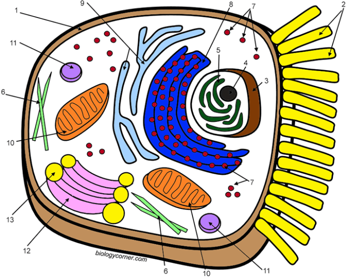
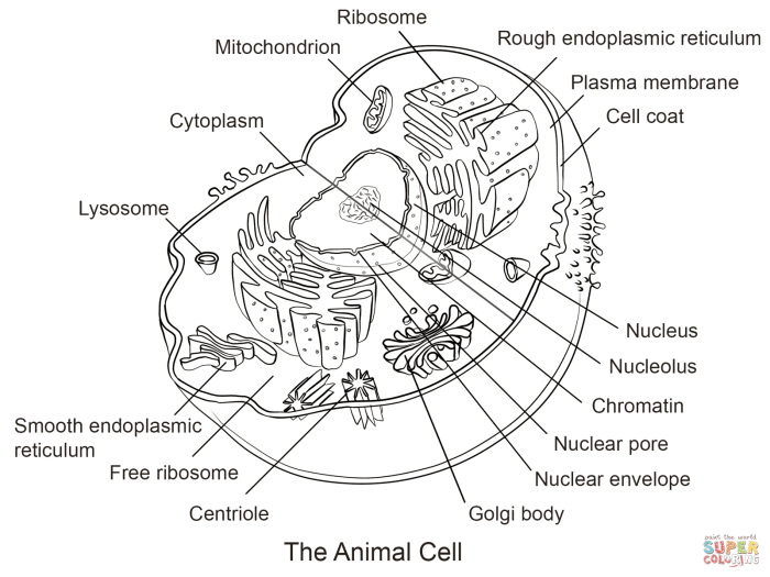
Animal cell with mitochondria coloring pages – Animal cells are the fundamental building blocks of animals, exhibiting a complex internal structure responsible for carrying out various life processes. Unlike plant cells, they lack a cell wall and chloroplasts. Instead, their defining features include a cell membrane, cytoplasm, and a variety of organelles, each with specific functions contributing to the overall health and operation of the cell.Mitochondria are essential organelles found within the cytoplasm of animal cells.
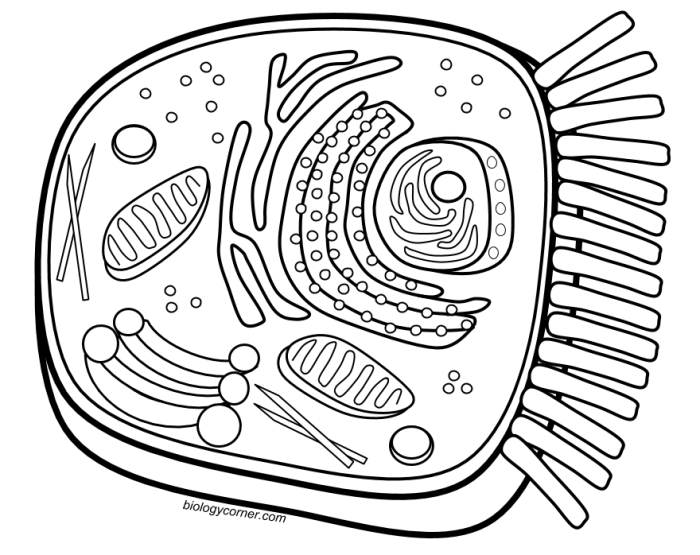
Animal cell with mitochondria coloring pages offer a fun way to learn about cellular biology. The intricate details of the mitochondria, the powerhouse of the cell, can be engaging for students of all ages. For a change of pace, you might also enjoy coloring some adorable animals, such as the ones found at animal beagle coloring pages , before returning to the fascinating world of cellular structures.
Understanding the mitochondria’s role within the animal cell is key to comprehending basic biological functions.
These structures are often referred to as the “powerhouses” of the cell because their primary function is to generate adenosine triphosphate (ATP), the cell’s primary energy currency. This energy is crucial for all cellular activities, from muscle contraction to protein synthesis and cell division. The efficiency of mitochondria directly impacts the overall health and function of the cell and the organism as a whole.
Mitochondrial Structure and Visual Representation
In a typical animal cell diagram, mitochondria are depicted as elongated, bean-shaped organelles. They possess a double membrane structure: an outer membrane that forms a smooth, continuous boundary and an inner membrane that is highly folded into cristae. These cristae significantly increase the surface area available for the electron transport chain, a crucial step in ATP production. The space between the inner and outer membranes is called the intermembrane space, while the space enclosed by the inner membrane is the mitochondrial matrix.
The matrix contains mitochondrial DNA (mtDNA), ribosomes, and enzymes involved in various metabolic processes. The visual representation often shows the inner membrane’s folded cristae as numerous internal ridges or folds within the bean-shaped structure, clearly differentiating it from the smooth outer membrane. The overall appearance is often depicted in varying shades of light brown or reddish-brown to distinguish it from other organelles.
The number of mitochondria within a cell can vary depending on the cell’s energy demands; highly active cells, such as muscle cells, typically contain a large number of mitochondria.
Designing a Coloring Page: Animal Cell With Mitochondria Coloring Pages
Creating an engaging and informative coloring page about animal cells and mitochondria requires careful consideration of layout and structure. The goal is to present complex biological information in a visually appealing and easily understandable manner for children. A well-designed coloring page can significantly enhance learning and retention.The arrangement of the cell’s components should be clear and logical, facilitating a child’s understanding of the relationships between different organelles.
The prominent placement of the mitochondria, highlighting their crucial role in energy production, is particularly important. Using a visually appealing color scheme and simple shapes will further enhance the learning experience.
Cell Component Placement and Visual Appeal
A successful coloring page layout balances visual appeal with educational accuracy. Consider using bright, engaging colors to differentiate organelles. Simple shapes and clear Artikels are crucial for easy coloring and understanding. Overly complex designs can be overwhelming for young children. The overall aesthetic should be inviting and encourage children to engage with the content.
The following table provides a sample layout for a four-column coloring page. Remember, this is just a suggestion; adjustments can be made based on the specific needs and age group of the intended audience.
| Nucleus (large, central circle with a smaller circle inside representing the nucleolus) | Cell Membrane (outermost boundary, a slightly wavy line) | Mitochondria (several bean-shaped structures scattered throughout the cell) | Golgi Apparatus (a stack of flattened sacs, near the nucleus) |
| Ribosomes (small dots scattered throughout the cytoplasm) | Endoplasmic Reticulum (a network of interconnected tubes and sacs, extending from the nucleus) | Lysosomes (small, circular structures scattered throughout the cytoplasm) | Cytoplasm (the space filling the cell, colored a lighter shade to differentiate from organelles) |
Illustrating the Mitochondria

Mitochondria are often depicted as the powerhouses of the cell, and accurately illustrating their structure is crucial for understanding their function. This section will guide you through representing the key features of a mitochondrion in your coloring page, ensuring an accurate and engaging visual representation.The mitochondrion’s unique structure is intimately linked to its role in cellular respiration. Understanding its internal components is essential for a biologically accurate depiction.
Mitochondrial Internal Structure
The mitochondrion possesses a double membrane system, creating distinct internal compartments. The outer membrane is smooth, while the inner membrane is extensively folded into structures called cristae. These cristae significantly increase the surface area available for the crucial reactions of cellular respiration. The space enclosed by the inner membrane is called the matrix. The matrix contains enzymes, ribosomes, and mitochondrial DNA (mtDNA), all necessary for energy production.
A visually accurate representation should clearly show the distinction between the outer and inner membranes, the intricate folding of the cristae, and the relatively homogenous appearance of the matrix. Imagine a bean-shaped structure with a highly folded inner membrane; that is a simplified visualization of the mitochondrion.
Mitochondrial Shape and Size
Mitochondria are typically depicted as bean-shaped or sausage-shaped organelles, although their morphology can be quite variable depending on the cell type and metabolic state. Their size is also not uniform, generally ranging from 0.5 to 10 micrometers in length. To illustrate their size relative to other organelles, consider including a scale bar on your coloring page, or representing them as noticeably larger than, say, ribosomes, which are much smaller, or roughly similar in size to the nucleus, which are much larger.
This comparison helps viewers grasp their relative dimensions within the cell.
Illustrating the Double Membrane
Imagine a drawing of a mitochondrion. The outer membrane should be depicted as a smooth, continuous line enclosing the entire organelle. Inside this outer membrane, the inner membrane is shown as a series of deeply folded, interconnected ridges – these are the cristae. The space between the outer and inner membranes is called the intermembrane space, which should be visually distinct.
The area enclosed by the inner membrane, filled with the matrix, should be clearly differentiated from the intermembrane space. The contrasting colors or shading of these three regions (outer membrane, intermembrane space, and inner membrane) would enhance the visual representation of this crucial double membrane structure. This detailed representation highlights the compartmentalization essential for the mitochondrion’s function.
Creating an Engaging Coloring Page Experience
Designing a captivating animal cell coloring page requires careful consideration of age appropriateness, clear labeling, and engaging accompanying instructions. The goal is to create a fun and educational activity that fosters understanding of cell structure. The coloring page should be visually appealing and accessible to a wide range of ages, from young children to older students.A well-designed coloring page should be adaptable to various age groups.
Younger children might benefit from larger, simpler Artikels of the cell’s main components, while older children can handle more detailed illustrations and finer lines. Consider incorporating different levels of complexity within the same page, perhaps with a simpler version overlaid on a more detailed one. This allows for flexibility and caters to individual skill levels.
Labeling the Cell Components
A clear and concise key is crucial for understanding the cell’s structure. Avoid overly technical terminology. Instead, use simple, descriptive labels that are easily understood by all ages. For instance, instead of “cytoplasm,” use “jelly-like substance filling the cell.” For the mitochondria, “powerhouses of the cell” is a memorable and informative label. The key should be visually appealing, perhaps using color-coded boxes matching the coloring instructions.
The key should be placed prominently on the page, possibly within a separate box or border.
Accompanying Instructions
Providing a set of clear and engaging instructions is key to enhancing the coloring experience. These instructions should guide the user through the coloring process step-by-step, making the activity accessible and enjoyable. The instructions should be presented in a visually appealing manner, perhaps using bullet points or a numbered list.
- Color the nucleus purple. This is the control center of the cell.
- Color the cell membrane light green. This is the outer boundary of the cell.
- Color the mitochondria blue. These are the energy producers of the cell.
- Color the endoplasmic reticulum (ER) yellow. It’s a network of membranes involved in protein synthesis.
- Color the Golgi apparatus orange. It packages and transports proteins.
- Color the lysosomes light brown. They are responsible for waste removal.
- Color the ribosomes dark green (small dots). They are involved in protein synthesis.
- Color the cytoplasm a pale yellow. This fills the cell.
Educational Value and Extensions

Coloring pages offer a unique and engaging approach to learning about complex biological concepts like animal cells and mitochondria. This hands-on activity transforms abstract ideas into visually appealing and memorable representations, making the learning process more enjoyable and effective, particularly for younger learners. The act of coloring encourages focus and attention to detail, reinforcing the understanding of the cell’s structure and the function of its components.The educational benefits extend beyond simple memorization.
The process of carefully coloring each part of the cell, such as the nucleus, cytoplasm, and, importantly, the mitochondria, helps students actively engage with the material, fostering a deeper understanding of their relative sizes and positions within the cell. This visual reinforcement aids retention and comprehension far beyond passively reading about these structures.
Extending the Learning Experience
Several activities can build upon the coloring page experience to further enhance learning and creativity. Creating a three-dimensional model of an animal cell, for example, allows students to apply their understanding of cell structure in a more tactile and interactive way. Students can use readily available materials such as clay, balloons, or even recycled materials to construct their models, further strengthening their grasp of the subject matter.
Researching specific aspects of mitochondria, such as their role in energy production or their involvement in diseases, can deepen their understanding of this crucial organelle. This research can take the form of short reports, presentations, or even collaborative projects, encouraging teamwork and information-sharing skills.
Fostering Creativity and Critical Thinking, Animal cell with mitochondria coloring pages
The coloring page can be a springboard for creative expression. Students are not limited to simply coloring within the lines; they can experiment with different color palettes, add details to their drawings, and even create their own unique artistic interpretations of the cell and its components. This freedom of expression encourages creativity and allows for individual expression of understanding.
Furthermore, the coloring page can be used to stimulate critical thinking. For instance, students could be challenged to design a coloring page showing a cell under different conditions (e.g., a healthy cell versus a cell experiencing energy depletion). This activity requires them to apply their knowledge and consider the impact of various factors on the cell’s structure and function.
It also encourages them to analyze and interpret information, key components of critical thinking.
Question Bank
What age group are these coloring pages suitable for?
These coloring pages can be adapted for various age groups. Younger children can focus on the basic shapes and colors, while older children can incorporate labeling and more detailed coloring.
Where can I find printable versions of these coloring pages?
Printable versions can be created from digital designs. Many online resources offer templates or allow for custom design and printing.
Are there any variations on this coloring page activity?
Yes, variations could include adding a 3D model creation component or researching specific mitochondrial diseases for older students.


