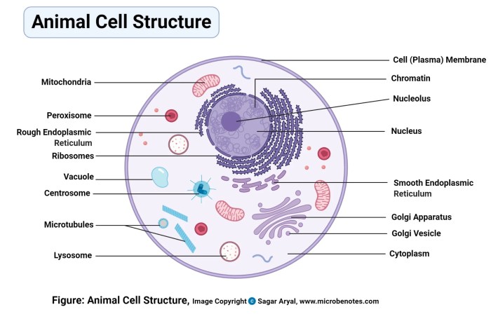
Common Color-Coding Schemes for Animal Cell Organelles

Animal cell coloring names – Color-coding is a crucial tool in visualizing and understanding the complex structures within an animal cell. Consistent color schemes aid in learning and memorization, making complex biological concepts more accessible. Different educational resources employ various color palettes, each with its own strengths and weaknesses. This section explores established conventions and proposes a novel scheme.
Examples of Established Color-Coding Conventions
Several established color-coding conventions exist for representing animal cell organelles in educational materials. These conventions often prioritize visual clarity and mnemonic associations to help students remember the function and location of each organelle. However, there is no single universally accepted standard. Variations in color choices arise due to personal preference, artistic license, and the specific focus of the educational resource.
Understanding animal cell coloring names, such as the nucleus or mitochondria, can be enhanced by visualizing their structures. A helpful resource for this is exploring the various shapes and forms through animal blank coloring pages , which provide a great foundation for understanding animal anatomy before diving into the complexities of cellular structures. This visual approach can aid in remembering the specific names and locations of the organelles within the animal cell.
Comparison of Three Different Color Schemes, Animal cell coloring names
Let’s compare three hypothetical color schemes. Scheme A might use blue for the nucleus (recalling its role in genetic information, often associated with the color of water, the source of life), red for mitochondria (representing their energy-producing function, often associated with heat and activity), and green for the chloroplast (if including it, a common mistake, as animal cells lack chloroplasts).
Scheme B might opt for a more pastel approach, using light yellow for the nucleus, light pink for the mitochondria, and light green for the endoplasmic reticulum. Scheme C could employ a vibrant palette, using bright orange for the Golgi apparatus, deep purple for lysosomes, and bright yellow for the nucleus. The effectiveness of each scheme depends on individual learning styles and the specific needs of the educational context.
For example, Scheme A’s strong, contrasting colors might be better for visual learners, while Scheme B’s softer colors might be less visually overwhelming. Scheme C’s brightness could be distracting for some.
A Novel Color-Coding Scheme
This section presents a new color-coding scheme designed for optimal visual clarity and mnemonic associations. The color choices are carefully selected to minimize confusion and aid in memorization.
| Organelle Name | Color | Justification | Mnemonic Device |
|---|---|---|---|
| Nucleus | Dark Blue | Represents the control center, evoking a sense of authority and stability. Darker shade provides contrast. | Blueprints (DNA housed within) |
| Mitochondria | Bright Red | Represents energy production, associating with heat and activity. Bright color for high energy. | Red Hot Energy |
| Endoplasmic Reticulum | Light Orange | A lighter shade to differentiate from the Golgi apparatus, but still warm and visually distinct. | Orange Road (transport network) |
| Golgi Apparatus | Dark Orange | A darker shade to distinguish it from the ER, representing its packaging and processing role. | Orange Packaging Plant |
| Lysosomes | Deep Purple | Represents the digestive function, suggesting mystery and breakdown. | Purple Power (waste disposal) |
| Ribosomes | Grey | A neutral color to represent their ubiquitous presence and fundamental role in protein synthesis. | Grey Builders (protein synthesis) |
| Cell Membrane | Light Green | Represents the boundary and protection, a soft color for a protective barrier. | Green Gatekeeper |
| Cytoplasm | Light Yellow | Represents the internal environment, a light and airy color for the internal fluid. | Yellow Interior |
Creative Color Choices and Their Rationale: Animal Cell Coloring Names
Choosing colors for representing organelles in an animal cell diagram goes beyond simple aesthetics; it significantly impacts understanding and retention. While traditional color-coding schemes exist, exploring unconventional palettes offers a fresh perspective, enhancing both visual appeal and cognitive engagement. The strategic use of color can leverage principles of color psychology to improve memorability and deepen comprehension of complex cellular structures.The benefits of using unconventional color choices are multifaceted.
Firstly, it combats the potential for rote memorization, encouraging a more active and analytical approach to learning. By breaking away from established norms, students are prompted to engage more deeply with the material, connecting color with function in a more meaningful way. Secondly, a unique color scheme can improve visual differentiation, particularly for organelles with subtle structural or functional differences that might be overlooked in a more standard representation.
Finally, a well-chosen palette can foster creativity and personalize the learning experience, making the study of cell biology more engaging and less daunting.
Color Psychology and Cell Structure Memorization
Color psychology plays a crucial role in how we perceive and remember information. Certain colors evoke specific emotions and associations, influencing our cognitive processing. For example, warm colors like reds and oranges are often associated with energy and excitement, while cooler colors like blues and greens are linked to calmness and stability. By strategically assigning colors to organelles based on their function, we can harness these associations to enhance memorization.
For instance, a vibrant red might represent the mitochondria, emphasizing their role as the powerhouse of the cell, while a serene blue could be used for the endoplasmic reticulum, highlighting its role in protein synthesis and cellular calm. This deliberate application of color psychology transforms the diagram into a more mnemonic device.
A Unique Visual Representation of an Animal Cell
Imagine an animal cell rendered with a unique color palette. The cell membrane is a deep, shimmering amethyst, reflecting its protective role and the intricate interactions it mediates. The nucleus, the control center, is a rich, warm ochre, suggesting its vital role in directing cellular processes. The mitochondria are depicted in a vibrant coral, representing their energetic activity and metabolic processes.
The rough endoplasmic reticulum is a deep teal, emphasizing its role in protein synthesis, while the smooth endoplasmic reticulum is a lighter, almost turquoise shade, reflecting its involvement in lipid metabolism. The Golgi apparatus, a crucial processing and packaging center, is rendered in a sophisticated burnt sienna, hinting at its complex functions. Lysosomes, the cellular recycling units, are depicted as a deep, almost black burgundy, signifying their role in waste breakdown.
The ribosomes, tiny protein factories, are represented as tiny, sparkling gold dots, highlighting their abundance and importance. The cytoskeleton, the cell’s structural support system, is visualized as a network of fine, silvery threads, suggesting its strength and flexibility. Finally, the cytoplasm, the jelly-like substance filling the cell, is a soft, translucent lavender, providing a calming background for the other organelles.
This palette moves beyond typical representations, creating a visually arresting and memorable image that links color to function in a creative and insightful way.
Coloring Activities and Educational Applications

Coloring activities offer a surprisingly effective and engaging method for learning about the intricate structures within an animal cell. The process of selecting colors and meticulously filling in the shapes of organelles helps solidify understanding in a way that passive reading or listening often cannot. This hands-on approach caters to various learning styles, enhancing memory retention and promoting a deeper comprehension of complex biological concepts.Coloring activities enhance understanding of animal cell structures by transforming abstract concepts into tangible, visual representations.
The act of associating specific colors with particular organelles creates a memorable link, improving recall and facilitating the organization of knowledge. Furthermore, the detailed nature of cell diagrams encourages close observation and attention to detail, fostering a more precise understanding of the relative sizes and positions of different organelles within the cell.
Step-by-Step Guide for an Animal Cell Organelle Coloring Activity
A well-structured coloring activity can significantly improve learning outcomes. The following steps Artikel a practical approach to guide students through the process:
- Introduction and Overview: Begin by providing a brief overview of animal cells and their key functions. Introduce the major organelles that will be colored, highlighting their roles within the cell. A short, engaging video or a simple diagram could be helpful here.
- Color Selection and Rationale: Distribute pre-printed diagrams of animal cells with clearly labeled organelles. Discuss the color-coding scheme chosen (e.g., nucleus – purple, mitochondria – red, etc.), explaining the rationale behind each color selection (e.g., purple for the nucleus to represent its importance as the control center; red for mitochondria to reflect their energy-producing role). Encourage students to consider the functions of each organelle when choosing their colors, if a free-coloring option is preferred.
- Coloring Process: Guide students through the coloring process, emphasizing neatness and accuracy in representing the shape and location of each organelle. Encourage them to refer back to the overview and color-coding scheme as needed.
- Labeling and Review: After coloring, have students label each organelle on their diagram. This reinforces the association between the visual representation and the organelle’s name. A brief review session, discussing the functions of each organelle, can further solidify their understanding.
- Assessment and Extension Activities: Assess student understanding through a short quiz or discussion. Extension activities could include creating a 3D model of an animal cell or researching specific organelles in more detail.
Catering to Various Learning Styles Through Coloring Activities
Different coloring activities can be adapted to suit diverse learning styles. For example, visual learners benefit from detailed, accurately labeled diagrams and a clear color-coding scheme. Kinesthetic learners may benefit from a more hands-on approach, perhaps creating a 3D model of the cell after completing the coloring activity. Auditory learners can benefit from verbal explanations and discussions about the functions of each organelle during the coloring process.
Providing varied approaches ensures that all learners can engage with the material effectively. For instance, a student might color a simplified cell diagram with only the major organelles first, and then progress to a more detailed diagram as their understanding improves. This layered approach allows for gradual skill development and caters to different levels of comprehension.
Top FAQs
What are some common mistakes in animal cell coloring?
Common mistakes include inconsistent color usage across diagrams, neglecting size ratios of organelles, and failing to clearly label structures.
How can I make my animal cell coloring more engaging for younger students?
Use bright, vibrant colors, incorporate fun shapes, and add simple details to make the organelles more relatable. Consider incorporating a story or game element.
Are there any online resources for printable animal cell coloring pages?
Many educational websites and online resources offer printable animal cell coloring pages with varying levels of detail and complexity. A quick internet search should yield several options.
Why is color-coding important in visualizing animal cells?
Color-coding helps differentiate organelles, making it easier to identify and remember their locations and functions. It significantly enhances visual comprehension and memorization.


