Introduction to Animal Eukaryotic Cells

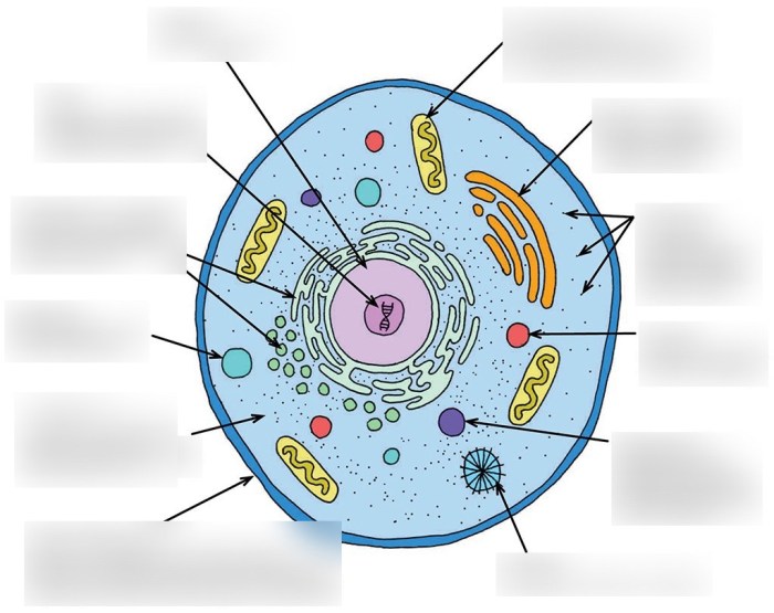
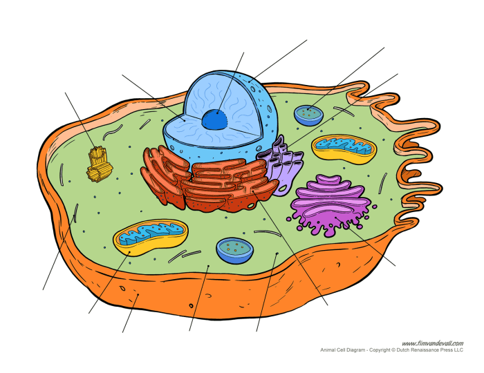
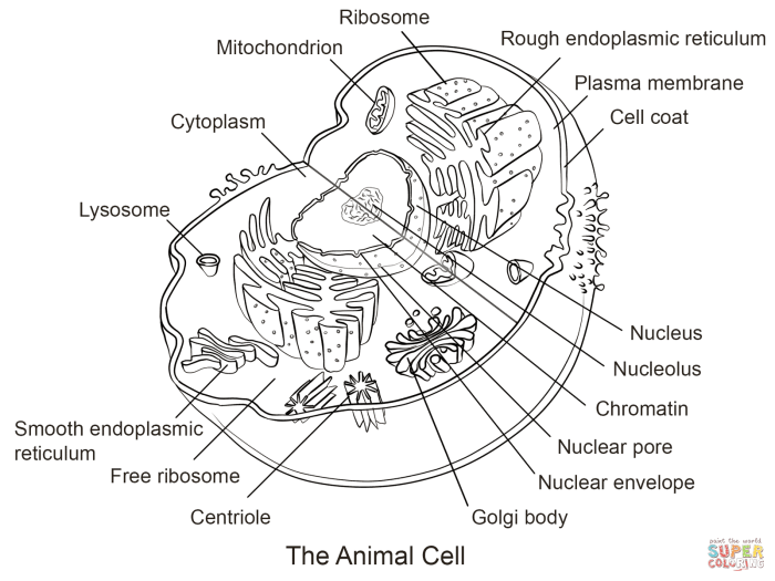
Animal eukaryotic cell coloring – Animal eukaryotic cells are the fundamental building blocks of animal tissues and organs. Unlike prokaryotic cells, they possess a complex internal structure characterized by membrane-bound organelles, each performing specialized functions crucial for the cell’s survival and overall organismal function. Understanding these organelles and their interactions is key to comprehending the intricacies of animal life.Animal eukaryotic cells are generally characterized by their possession of a membrane-bound nucleus, housing the cell’s genetic material, and numerous other organelles working in coordination.
These organelles compartmentalize cellular processes, increasing efficiency and reducing the likelihood of detrimental interactions between different biochemical pathways.
The Cell Membrane, Animal eukaryotic cell coloring
The cell membrane, also known as the plasma membrane, is a selectively permeable barrier that encloses the cell’s cytoplasm and organelles. It’s primarily composed of a phospholipid bilayer, with hydrophilic phosphate heads facing outwards towards the aqueous environments (intracellular and extracellular fluids) and hydrophobic fatty acid tails oriented inwards. Embedded within this bilayer are various proteins, cholesterol molecules, and glycolipids.
These components contribute to the membrane’s fluidity, selective permeability, and ability to interact with its surroundings. The cell membrane regulates the passage of substances into and out of the cell through various mechanisms, including passive diffusion, facilitated diffusion, active transport, and endocytosis/exocytosis. This control is essential for maintaining the cell’s internal environment and carrying out its functions.
The Nucleus
The nucleus is the cell’s control center, housing the genetic material in the form of DNA organized into chromosomes. The nuclear envelope, a double membrane studded with nuclear pores, regulates the transport of molecules between the nucleus and the cytoplasm. Within the nucleus, the nucleolus is responsible for ribosome biogenesis. The nucleus dictates the cell’s activities by controlling gene expression and regulating protein synthesis.
Mitochondria
Mitochondria are often referred to as the “powerhouses” of the cell. These double-membrane bound organelles are the sites of cellular respiration, where glucose is broken down to produce ATP (adenosine triphosphate), the cell’s primary energy currency. The inner membrane of the mitochondria is folded into cristae, significantly increasing its surface area and maximizing ATP production. Mitochondria also play a role in other cellular processes, including calcium homeostasis and apoptosis (programmed cell death).
Ribosomes
Ribosomes are small, RNA-protein complexes responsible for protein synthesis. They are found free in the cytoplasm or bound to the endoplasmic reticulum. Free ribosomes synthesize proteins for use within the cytoplasm, while bound ribosomes produce proteins destined for secretion or insertion into membranes. The process of protein synthesis, or translation, involves the decoding of mRNA (messenger RNA) sequences to assemble amino acids into polypeptide chains.
Endoplasmic Reticulum
The endoplasmic reticulum (ER) is an extensive network of interconnected membranes extending throughout the cytoplasm. There are two main types: rough ER (RER) and smooth ER (SER). The RER is studded with ribosomes, giving it its rough appearance. It’s involved in protein synthesis, folding, and modification. The SER lacks ribosomes and plays roles in lipid synthesis, detoxification, and calcium storage.
Golgi Apparatus
The Golgi apparatus, also known as the Golgi complex or Golgi body, is a stack of flattened, membrane-bound sacs (cisternae). It receives proteins and lipids from the ER, further modifies them (e.g., glycosylation), sorts them, and packages them into vesicles for transport to their final destinations – either within the cell or for secretion outside the cell. The Golgi apparatus acts as a central processing and distribution hub for cellular materials.
Lysosomes
Lysosomes are membrane-bound organelles containing hydrolytic enzymes that break down various cellular waste products, including damaged organelles and ingested materials. They maintain cellular health by recycling cellular components and protecting against pathogens. The acidic environment within lysosomes optimizes the activity of these hydrolytic enzymes.
Interpreting Stained Cell Images

Microscopic examination of stained animal eukaryotic cells is crucial for understanding their structure and function. Different stains bind to specific cellular components, revealing their location, shape, and relative abundance. Analyzing the color and intensity of these stains allows researchers to infer the cell’s health and activity.Staining techniques provide a visual representation of otherwise invisible cellular structures. The intensity of the stain reflects the concentration of the targeted molecule within the cell, offering insights into cellular processes.
Variations in staining patterns can indicate cellular abnormalities, such as damage or disease.
Examples of Stained Animal Eukaryotic Cells
Imagine viewing a slide under a microscope. We’ll consider three hypothetical examples to illustrate the principle of interpreting stained cell images. These examples are simplified for clarity but represent the basic principles.First, consider a cell stained with hematoxylin and eosin (H&E), a common stain in histology. The hematoxylin, a basic dye, stains the nucleus a deep purple or blue, highlighting the densely packed chromatin.
The eosin, an acidic dye, stains the cytoplasm a pale pink, revealing its texture and the presence of any cytoplasmic inclusions. A healthy cell would show a clearly defined, centrally located nucleus with evenly stained chromatin and a uniformly pink cytoplasm.Second, consider a cell stained with a fluorescent dye specific to microtubules, such as a dye conjugated to an antibody against tubulin.
Under fluorescence microscopy, the microtubules would appear as bright green filaments radiating from a central organizing center (the centrosome), showcasing their organization within the cell’s cytoskeleton. The intensity of the green fluorescence would indicate the abundance of microtubules, which is related to the cell’s ability to maintain its shape and transport organelles. A disrupted or disorganized microtubule network might suggest cellular stress or dysfunction.Third, let’s visualize a cell stained with a dye specific to mitochondria, perhaps a fluorescent dye that accumulates in the mitochondria’s membrane potential.
Healthy mitochondria would appear as numerous, bright red puncta (small dots) distributed throughout the cytoplasm. A cell with damaged or dysfunctional mitochondria might show fewer or fainter red puncta, indicating impaired energy production. The distribution of the staining could also be altered, showing clustering or uneven distribution.
Interpreting Staining Intensity and Color
The color intensity of a stain is directly proportional to the concentration of the targeted molecule. A deeply stained nucleus, for example, indicates a high concentration of DNA, potentially reflecting a cell actively engaged in transcription. Conversely, a faintly stained nucleus might suggest a cell that is less metabolically active. Similarly, intensely stained mitochondria would indicate high energy demands, whereas weak staining might indicate energy deficiency.Variations in staining patterns can indicate cellular dysfunction.
Understanding animal eukaryotic cell coloring involves appreciating the intricate details of organelles. For a simpler, visual introduction to animal structures, you might find resources like this helpful: animal coloring sheet free. These sheets offer a fun way to grasp basic animal anatomy before delving into the complexities of cellular structures and their respective coloring patterns in microscopic study.
For example, uneven staining of the nucleus might suggest chromatin damage or abnormal gene expression. Fragmented or condensed chromatin, appearing as dark, irregular clumps, can be indicative of apoptosis (programmed cell death). Irregular staining patterns in the cytoplasm might indicate the presence of abnormal inclusions or organelles.
Healthy versus Unhealthy Cells: Staining Patterns
A healthy cell typically exhibits uniform staining patterns, with clearly defined organelles and a consistent intensity of staining across different cellular components. In contrast, an unhealthy cell might show irregular staining, with variations in intensity and distribution. For instance, a cell undergoing apoptosis might show condensed and fragmented chromatin, along with cytoplasmic blebbing (formation of membrane-bound protrusions). Cells with damaged organelles, such as mitochondria, might exhibit altered staining patterns within these organelles.
In summary, consistent and uniform staining generally indicates cellular health, while irregular staining patterns often signal cellular stress, damage, or disease.
Applications of Cell Coloring Techniques: Animal Eukaryotic Cell Coloring

Cell staining techniques are indispensable tools in various biological disciplines, providing crucial insights into cellular structure, function, and pathology. These techniques allow researchers and clinicians to visualize specific cellular components, identify abnormalities, and ultimately contribute to accurate diagnoses and effective treatments. The applications are diverse and far-reaching, impacting our understanding of both healthy and diseased tissues.The ability to visualize cellular components with specific stains allows for detailed analysis of cellular morphology, identification of specific organelles, and the detection of abnormal cellular processes.
This information is critical in various fields, from basic research to clinical diagnostics. The choice of stain depends heavily on the specific target and the information desired.
Applications in Histology, Cytology, and Pathology
Histological, cytological, and pathological analyses rely heavily on cell staining to reveal the microscopic architecture of tissues and cells. Histology focuses on the microscopic examination of tissues, cytology on individual cells, and pathology on the nature and cause of diseases. These fields utilize a range of staining methods to highlight different cellular components and identify disease markers.
- Hematoxylin and Eosin (H&E) staining: This is a routine stain used in histology and pathology to visualize the nuclei (hematoxylin, stains blue/purple) and cytoplasm (eosin, stains pink/red) of cells. This provides a general overview of tissue architecture and cellular morphology, enabling the identification of various tissue types and the detection of abnormalities like inflammation or tumors.
- Periodic acid-Schiff (PAS) staining: PAS stain is used to detect polysaccharides and glycoproteins, which are abundant in connective tissues and some microorganisms. It’s particularly useful in identifying fungal infections and glycogen storage diseases. The stain produces a magenta color in positive areas.
- Immunohistochemistry (IHC): IHC uses antibodies to target specific proteins within cells. This technique is incredibly powerful for identifying specific cell types, diagnosing cancers (e.g., identifying specific cancer markers), and studying the expression of proteins involved in various cellular processes. The results can be visualized using chromogenic or fluorescent detection methods.
Disease Diagnosis and Treatment
Cell staining techniques are crucial for the diagnosis and monitoring of various diseases. The ability to visualize specific cellular features or markers allows for early detection, accurate classification, and effective treatment strategies.
- Cancer diagnosis: IHC is extensively used in cancer diagnosis to identify specific tumor markers, assess tumor grade, and predict prognosis. For example, staining for ER (estrogen receptor), PR (progesterone receptor), and HER2 receptors in breast cancer helps determine the appropriate treatment strategy. Positive staining indicates hormone receptor-positive breast cancer, which is treated differently than hormone receptor-negative breast cancer.
- Infectious disease diagnosis: Staining techniques, such as Gram staining for bacteria and PAS staining for fungi, are essential for identifying infectious agents in clinical samples. This rapid identification is crucial for guiding appropriate antibiotic or antifungal therapy.
- Neurological disorders: Specific stains can help diagnose neurological disorders by visualizing the presence of amyloid plaques (in Alzheimer’s disease) or tau tangles (in various neurodegenerative diseases). These techniques aid in confirming diagnoses and understanding disease progression.
Top FAQs
What are the safety precautions when working with cell stains?
Always wear appropriate personal protective equipment (PPE), including gloves and eye protection, when handling stains. Many stains are toxic or irritants. Work in a well-ventilated area and follow the manufacturer’s safety guidelines.
How can I determine the optimal staining time for my experiment?
Optimal staining time varies depending on the specific stain and the type of cells being studied. Pilot experiments are often necessary to determine the ideal staining duration, ensuring adequate staining while minimizing overstaining artifacts.
What are some common artifacts that can occur during cell staining?
Common artifacts include precipitate formation from the stain, uneven staining due to inadequate sample preparation, and damage to the cells during the staining process. Careful technique and proper controls are essential to minimize artifacts.


