Introduction to Animal Cell Structure

Biologycomer.com animal cell coloring – Animal cells are the fundamental building blocks of animal tissues and organs. Unlike plant cells, they lack a rigid cell wall and chloroplasts, resulting in a more flexible and diverse range of shapes and sizes. Understanding their intricate structure is crucial to comprehending the complexities of animal life and physiology. This section will explore the key components of animal cells and their respective functions, highlighting the differences between animal and plant cells, and providing a detailed look at the cell membrane.Animal cells contain a variety of organelles, each with a specific role in maintaining cellular function.
These organelles work together in a coordinated manner to ensure the cell’s survival and contribute to the overall functioning of the organism. Key differences exist between animal and plant cells, primarily concerning the presence or absence of certain organelles and structural features.
Animal Cell Components and Their Functions
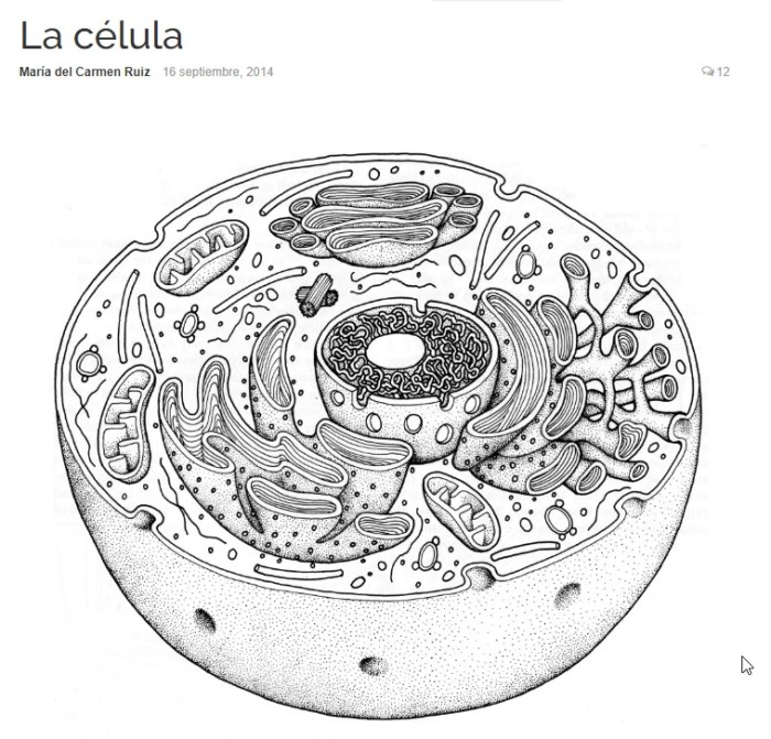
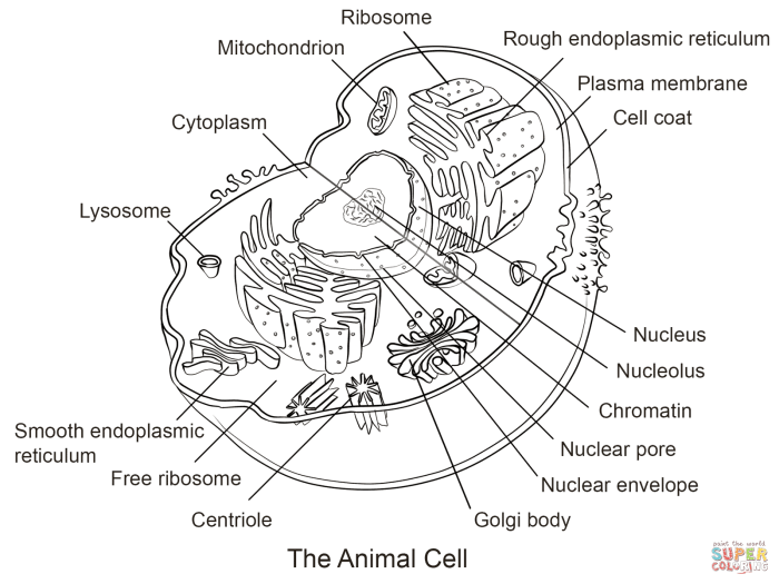
The cytoplasm, a gel-like substance filling the cell, houses various organelles. The nucleus, often considered the cell’s control center, contains the genetic material (DNA) organized into chromosomes. Ribosomes, responsible for protein synthesis, are found free-floating in the cytoplasm or attached to the endoplasmic reticulum (ER). The ER, a network of membranes, plays a critical role in protein and lipid synthesis and transport.
The smooth ER synthesizes lipids, while the rough ER, studded with ribosomes, modifies and transports proteins. The Golgi apparatus further processes and packages proteins for secretion or use within the cell. Mitochondria, often referred to as the “powerhouses” of the cell, generate energy in the form of ATP through cellular respiration. Lysosomes contain enzymes that break down waste materials and cellular debris.
The cytoskeleton, a network of protein filaments, provides structural support and facilitates cell movement.
Differences Between Plant and Animal Cells
A significant difference between plant and animal cells lies in the presence of a cell wall in plant cells. This rigid outer layer provides structural support and protection, absent in animal cells. Plant cells also possess chloroplasts, organelles responsible for photosynthesis, which are not found in animal cells. Plant cells typically have a large central vacuole for storage and maintaining turgor pressure, whereas animal cells may have smaller vacuoles or lack them altogether.
These structural differences reflect the distinct physiological needs and functions of plant and animal cells.
The Cell Membrane and Its Role
The cell membrane, also known as the plasma membrane, is a selectively permeable barrier that encloses the cytoplasm and regulates the passage of substances into and out of the cell. It is composed primarily of a phospholipid bilayer, with embedded proteins. The phospholipid bilayer consists of two layers of phospholipid molecules, each with a hydrophilic (water-loving) head and two hydrophobic (water-fearing) tails.
This arrangement creates a barrier that prevents the free passage of most molecules. Membrane proteins perform various functions, including transport of molecules, cell signaling, and cell adhesion. The fluid mosaic model describes the dynamic nature of the membrane, where components are constantly moving and interacting. The selective permeability of the cell membrane is crucial for maintaining homeostasis within the cell, ensuring the proper balance of ions and molecules necessary for cellular processes.
This selective permeability is achieved through various mechanisms, including passive transport (diffusion, osmosis) and active transport (requiring energy).
Coloring Activity Design: Biologycomer.com Animal Cell Coloring

This section details the design of a simplified animal cell diagram suitable for a coloring activity, focusing on key organelles and their representative colors. The worksheet aims to provide a visually engaging and educational experience for students learning about animal cell structure. The design prioritizes clarity and ease of understanding, ensuring that the coloring activity reinforces learning rather than adding complexity.
The worksheet will present a large, central diagram of an animal cell, simplified to include only the most essential organelles. Each organelle will be clearly labeled and Artikeld, making it easy for students to color them according to the provided key. The overall design will be visually appealing, utilizing clear lines and ample space to prevent overcrowding. The use of color will aid in differentiating between the various organelles and their functions.
Organelle Selection and Color Assignment, Biologycomer.com animal cell coloring
The following organelles will be included in the simplified animal cell diagram, each assigned a distinct color to enhance visual learning and memorization. The color choices are based on common visual representations found in educational materials, ensuring consistency and ease of understanding. These colors are suggestions and can be adapted based on available coloring materials.
The selection of these five organelles provides a representative overview of the major components and functions of an animal cell. Students will be able to visualize the relative positions of these structures and associate them with their respective roles in cellular processes.
- Nucleus: Light Purple. The nucleus, the control center of the cell, will be depicted as a large, centrally located structure.
- Cell Membrane: Light Blue. The cell membrane, the outer boundary of the cell, will be represented as a thin, enclosing layer.
- Mitochondria: Dark Red. Mitochondria, the powerhouses of the cell, will be shown as numerous, elongated structures scattered throughout the cytoplasm.
- Ribosomes: Dark Green. Ribosomes, responsible for protein synthesis, will be depicted as small, numerous dots scattered throughout the cytoplasm.
- Golgi Apparatus: Light Green. The Golgi apparatus, involved in packaging and transporting proteins, will be shown as a series of flattened sacs.
Organelle Key
The following table provides a key identifying each organelle and its assigned color. This key will be included on the worksheet to aid students in completing the coloring activity accurately. The table’s responsive design ensures readability across various devices.
| Organelle Name | Color |
|---|---|
| Nucleus | Light Purple |
| Cell Membrane | Light Blue |
| Mitochondria | Dark Red |
| Ribosomes | Dark Green |
| Golgi Apparatus | Light Green |


